New technique for 3D cell imaging developed
A high-throughput method of imaging biological particles using an X-ray laser has been developed by an international team of scientists led by Uppsala University, Sweden.
The experiment, which represents a major milestone for studies of individual biological structures using X-ray lasers, has provided images of projections of the carboxysome particle, a delicate and tiny cell compartment in photosynthetic bacteria.
The technique paves the way for 3D imaging of parts of the cell, and even small viruses, to develop a deeper understanding of life’s smallest units.
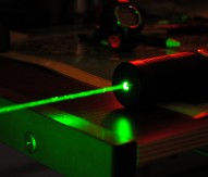
Using a specially designed injector that produces a particle stream thinner than a human hair, the scientists sprayed an aerosol of carboxysomes – a cell organelle for CO2 assimilation in cyanobacteria and which contain protein machinery that incorporates carbon from carbon dioxide into biomolecules – across the beam of the LCLS X-ray laser at the SLAC National Accelerator Laboratory in the US.
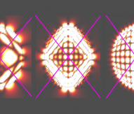
The scientists calculated the structure of the organelles by analysing the way the carboxysomes scatter the extremely short and ultra bright X-ray flashes of the LCLS. Until now, this method required crystals of the sample material to get sufficient signal. Thanks to the extreme brightness of the X-ray laser and clever analysis of the diffraction patterns, the researchers could reconstruct individual samples without having to crystallise them.
Carboxysomes like many other biological samples vary in shape and size and therefore cannot be crystallised.
Within minutes, the researchers collected 70,000 scattering patterns from individual particles. The analysis returned an icosahedral shape (a structure with 20 triangle-shaped sides) for carboxysomes, in line with expectations. The results also showed considerable variation in size.
“Our method allows single-particle imaging of objects which can be different in size and shape”, said Max Hantke, a doctoral student in molecular biophysics at Uppsala University, who led the research.
He added: “With the carboxysomes we have reconstructed the smallest single biological particles ever imaged with an X-ray laser, and we were also able to improve resolution. The reconstruction shows details as small as about 18 nanometres. For the first time we access a very interesting size regime with an X-ray laser. Large pathogenic viruses like HIV, influenza, and the herpes virus are in the same size domain as the carboxysome.”
Hantke’s supervisor, Filipe Maia, concluded: “These results show the way to high-throughput imaging of biological samples at high resolution. High data rates and very short exposures allow studies on the dynamics of particles and permit the analysis of structural variations, which are crucially important for life.”
The research has been published in the journal Nature Photonics.